Article Text

Abstract
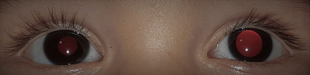
Case presentation A 10-month-old boy was admitted to the emergency department due to a sudden onset of left unilateral mydriasis (figure 1). His medical history was unremarkable. A minor head trauma 2 days before was reported, without alarming signs or symptoms. His mother was putting him to sleep, after coming back from work, when she noticed a different pupil size and promptly went to the ED with her husband. The parents denied any use of medications, including nebulised therapy or direct contact with plants. The child was well appearing and his vital signs were within the standard age limits. His extraocular motility was normal as well as the rest of his neurological and physical examination. Parents’ behaviour was somehow remarkable. Even though the child was not suffering, the mother seemed very worried while the father was nervous and aggressive, repeatedly asking for a discharge.
Left unilateral mydriasis.
Questions
What is the most likely diagnosis based on this clinical presentation?
Local contact with a mydriatic substance
Intracerebral haemorrhage
Brain tumour
Third nerve palsy
What would be the next step in the investigation to confirm this diagnosis?
Brain CT
Brain MRI
Fundus oculi examination
Toxicological screening of urine
How is this condition managed, and what is the prognosis?
Questions Answers can be found on page XX
- toxicology
- ophthalmology
- child abuse
Statistics from Altmetric.com
Footnotes
Contributors AGS, FP and EB conceptualised and drafted the initial manuscript and reviewed and revised the manuscript. RD, LD, AT and LC designed data collection instruments, coordinated and supervised data collection, and critically reviewed the manuscript. All authors approved the final manuscript as submitted and agree to be accountable for all aspects of the work.
Funding The authors have not declared a specific grant for this research from any funding agency in the public, commercial or not-for-profit sectors.
Competing interests None declared.
Provenance and peer review Not commissioned; externally peer reviewed.