Article Text

Abstract
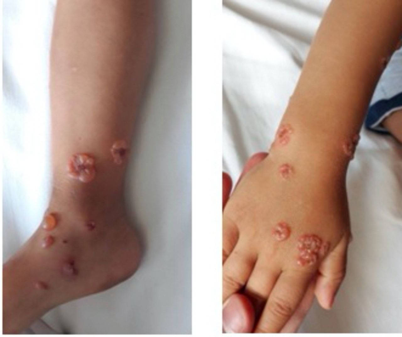
A 3-year-old boy presented with a 5-day history of bullous skin lesions localised mainly in the upper and lower limbs and in the genital region (figure 1). Lesions were not pruritic nor painful and showed a central crust. There was no family history of skin disorders or autoimmune diseases. The child never had fever and his physical examination was otherwise unremarkable.

Bullous skin lesions forming around a central crust, localised in the upper and lower limbs.
Questions
What is the most likely diagnosis based on this clinical presentation?
Bullous impetigo.
Bullous pemphigoid.
Linear IgA bullous dermatosis.
Dermatitis herpetiformis.
What would be the next step in the investigation to confirm your diagnosis?
Skin biopsy.
Swab test for bacterial culture with an antibiogram.
Anti-transglutaminase antibody detection.
What is the mainstay of management?
Dapsone.
Systemic steroids.
Topical steroids.
All of the above answers are correct, according to the severity of the disease.
Questions Answers can be found on page 02 .