Article Text

Download PDFPDF
A strident girl
  1. Francesca Corrias1,
  2. Valentina Gesuete2,
  3. Flora Maria Murru3,
  4. Massimo Maschio4,
  5. Egidio Barbi4,5
  1. 1 Pediatric Department, University of Trieste, Trieste, Italy
  2. 2 Cardiology Service, IRCCS Materno Infantile Burlo Garofolo, Trieste, Italy
  3. 3 Radiology Service, IRCCS materno infantile Burlo Garofolo, Trieste, Italy
  4. 4 IRCCS Materno Infantile Burlo Garofolo, Trieste, Italy
  5. 5 University of Trieste, Trieste, Italy
  1. Correspondence to Dr Valentina Gesuete, Cardiology Service, IRCCS Materno Infantile Burlo Garofolo, Trieste 34134, Italy; valegesuete{at}gmail.com

Abstract

A 12-year-old girl was referred with a 7-month history of episodes of dyspnoea, stridor and a sense of chest constriction during physical exercise. These were self-limiting and never occurred at night. Physical examination was normal. An initial diagnosis of vocal cord dysfunction was made.

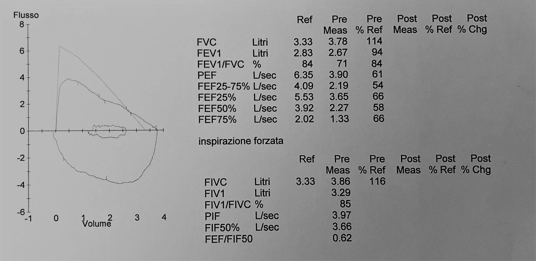
Spirometry showed a plateau of forced expiratory flow, with a truncated aspect of the expiratory phase (figure 1). Six weeks later she described occasional dysphagia with solid foods. The barium swallow, performed only in anteroposterior projection, did not show oesophageal dilation. A bronchoscopy showed extrinsic compression of the middle lower third of the trachea and the right main bronchus. A chest CT scan was performed (figures 2 and 3).

Figure 1 The spirometry showed a truncated expiratory phase with a substantially decreased PEF, compared with FEV1: indicating central intrathoracic airway obstruction. FEF, forced expiratory flow; FEV1, forced expiratory volume in 1 s; FIF, forced inspiratory flow; FIV1, forced inspiratory volume in 1 s; FIVC, forced inspiratory vital capacity; FVC, forced vital capacity; PEF, peak expiratory flow; PIF, peak inspiratory flow.

Figure 2 Contrast enhanced CT axial section image showing right aortic arch (white arrow on the left) with aberrant subclavian artery (red arrow on the right) encircling the trachea and the oesophagus.

Figure 3 CT three-dimensional reconstruction arteriography posterior view showing right aortic arch (white arrow), diverticulum of Kommerell (white star) from which the left subclavian artery (red arrow) arose. D Ao, descending aorta.

Questions

  1. What is your diagnosis?

    1. Persistent vocal cord dysfunction

    2. Achalasia

    3. Vascular ring

    4. Asthma

  2. What is the gold standard for diagnosis of VR?

    1. ECG

    2. Chest radiograph

    3. CT and/or MRI

    4. Bronchoscopy

  3. How should this patient be treated?

    1. Surgical correction

    2. Video-assisted thoracoscopy

    3. Decompression of the oesophagus with a nasogastric tube

    4. Inspiratory muscle training and ipratropium bromide inhaler

  4. What signs in the history pointed away from vocal cord dysfunction?

    1. Dysphagia with solid food was present.

    2. The episodes of dyspnoea and stridor never occurred at night.

    3. The episodes arose mainly on exertion.

    4. The episodes of dyspnoea and stridor were self-limiting.

Questions Answers can be found on page 2.

  • congenital heart disease
  • paediatric cardiac surgery
  • vascular ring

Statistics from Altmetric.com

Request Permissions

If you wish to reuse any or all of this article please use the link below which will take you to the Copyright Clearance Center’s RightsLink service. You will be able to get a quick price and instant permission to reuse the content in many different ways.

Footnotes

  • Competing interests None declared.

  • Provenance and peer review Not commissioned; externally peer reviewed.