Article Text

Abstract
Introduction Pierre Robin sequence (PRS) is a congenital anomaly presenting with micrognathia, glossoptosis and a cleft palate. This study describes a decade's experience of the management of upper airway obstruction (UAO) in PRS patients with a nasopharyngeal airway (NPA).
Methods This study was conducted by paediatric respiratory and otolaryngology departments. Children with PRS referred with UAO were evaluated according to a standard protocol. Data collected included the degree of airway obstruction, method of airway management, polysomnography data before and after intervention, and longer term follow-up.
Results Data were collected on 104 PRS patients referred to us for airway assessment in 2000–2010. 64/104 were aged <4 weeks at referral. Airway symptoms were managed conservatively in 27 patients (25.9%), with an NPA in 63 (60.6%) and a tracheostomy in 14 (13.4%). The average duration of NPA use was 8 months (3 weeks to 27 months). Polysomnography results improved in all 63 patients with an NPA. Fourteen severely obstructed patients underwent a tracheostomy. 86.5% (90/104) of PRS patients were managed conservatively or with the help of an NPA. There were no NPA related complications.
Conclusion There is a spectrum of UAO in PRS. This study reports on long-term outcomes in 104 children with PRS and airway obstruction. In most children (86.5%), airway obstruction was managed by conservative measures or with an NPA for a few months. The natural history shows that with normal growth, airway compromise resolves without immediate surgical intervention as advocated by some practitioners. Few PRS children require a tracheostomy.
Statistics from Altmetric.com
Introduction
Pierre Robin sequence (PRS) is a congenital anomaly that includes micrognathia, glossoptosis and a cleft palate.1 The incidence is approximately 1 in 85 000 children and can be seen either as an isolated sequence or in association with other congenital defects.2 These children often present with obstructed upper airways, stridor, cyanotic episodes and feeding difficulty. There have been many reports on different strategies to manage the airway in these patients,3 4 but no consensus has been reached. The objective of this study was to present our experience of airway management in PRS patients with special reference to the role of the nasopharyngeal airway (NPA).
What is already known on this topic
▶ Pierre Robin sequence (PRS) is a congenital anomaly presenting with micrognathia, glossoptosis and cleft palate.
▶ There have been previous reports on different strategies to manage upper airway obstruction in PRS patients.
What this study adds
▶ This is the first reported study on the long-term outcomes of 104 children with PRS and airway obstruction.
▶ Our report is the first to evaluate the nasopharyngeal airway (NPA) in a large PRS cohort managed over 10 years.
▶ In most children with PRS, airway obstruction can be managed by conservative measures or with an NPA.
Methods
This study was conducted jointly by the paediatric respiratory and otolaryngology departments at Great Ormond Street Hospital for Children (GOSH), London. All children with the full PRS of all three characteristics (micrognathia, glossoptosis and cleft palate) referred to GOSH from January 2000 to December 2010 were included. This study was a retrospective review of the patient records. The data collected included patient demographics, age at presentation, mode of airway management and serial sleep study data before and after intervention. The duration of use of the NPA and any associated complications and failures were evaluated. Data were also collected on feeding management.
For the study period, a standard protocol (see online supplementary flowchart 1) for managing upper airway obstruction (UAO) in PRS was followed. The children were admitted to the respiratory ward for airway assessment and an overnight sleep study was carried out, which included essential clinical observations by experienced staff. For the purpose of this study, the sleep studies were classified as absent/mild/moderate or severe UAO using the criteria described by Nixon et al.5 Broadly, the sleep study was classified as mild UAO if there was a set of at least three clusters of desaturations with at least 3 dips below 90% (but not below 85%), moderate UAO for a set of at least three clusters of desaturations with at least 3 dips below 85% (but not below 80%) and severe UAO for a set of at least three clusters of desaturations with at least 3 dips below 80%. Clinical observations by experienced staff were also taken into account, that is, mild UAO could be reclassified as moderate if staff reported a lot of obstruction events or moderate UAO could be reclassified as mild if the child had no respiratory effort (indicating desaturations were caused by central events). The objective oxygen saturation findings usually correlated with clinical observations. In only 5% of cases was the classification altered on the basis of clinical findings. If the sleep study demonstrated mild UAO, the patient underwent a trial of positioning, feeding and reflux management. If the sleep study indicated moderate or severe obstruction, an NPA was trialled. If the child was clinically severely obstructed on admission, an NPA was inserted without a pre-intervention sleep study.
A follow-up sleep study was carried out with the NPA in situ in all patients. If the NPA relieved the airway obstruction, the parents were trained to manage the NPA at home. At this stage, feeding was also reviewed. Once the parents were deemed competent to manage the NPA, the child was discharged home with replacement NPAs and suction apparatus. Parents were also taught how to recognise NPA blockade (inability to suction/reduced airflow) and change the NPA when blocked or routinely at 4–6-week intervals. Follow-up sleep studies were undertaken at 2-month intervals. A trial of NPA removal was scheduled according to clinical and sleep study results. After NPA removal, a confirmatory sleep study was carried out.
The NPAs used in these patients were custom made using an endotracheal tube (figure 1A) cut to length for the individual patient. The shortened endotracheal tube was then sutured to a tube holder and the NPA was ready for use (figure 1B). Correct positioning of the tip of the NPA just beyond the base of the tongue but above the epiglottis was confirmed using a lateral neck radiograph.

Nasopharyngeal airway consisting of a shortened endotracheal tube and a tube holder (A) sutured together ready for use (B).
For those patients with an NPA in situ who still had severe UAO, different sizes and lengths of NPA were assessed. If the airway obstruction was not relieved after a satisfactory trial of different NPAs, the child was referred to ENT surgeons for a microlaryngobronchoscopy and consideration for tracheostomy.
Results
In this series, 104 patients (48 female, 56 male) with all three PRS characteristics were referred for airway assessment. Age at the time of referral varied from 1 day to 12 months. A large number of patients (64/104) were less than 1 month of age at referral, with 48 of these 64 being less than 2 weeks of age. The majority of the patients (94/104) were born at term.
Of the 104 patients, 86 had PRS as their only abnormality. However, associated or additional diagnoses were noted in a further 18 children: Sticklers syndrome (five), Weissenbacher syndrome (one), Dubowitz syndrome (one), Van der Woude syndrome (one), Wolf Hirschhorn syndrome (one), trisomy 13 (one), 22/11 translocation (one), del 4q33 (one), Blackfan Diamond anaemia (one), trisomy 21 (one), exomphalos (one), 17q21 deletion (one), cystic fibrosis (one) and hydronephrosis (one).
The distribution of the cohort classified according to the airway management algorithm is summarised in online supplementary flowchart 2. The UAO was reported as mild on the initial sleep study in 27 patients. For these 27 patients (25.9%), the airway was successfully managed conservatively through positioning. These patients were observed clinically while awake, asleep and feeding before discharge and with sleep studies at regular intervals to assess their progress.
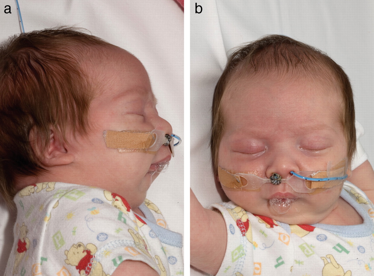
A total of 77 patients had moderate or severe UAO and underwent initial treatment with an NPA, which was successful in 63/77 (81.8%). The outer diameter of the endotracheal tube in this group varied from 2.5 to 3.5 mm and the length varied from 6.5 to 9 cm. Figure 2A,B shows a child with PRS and an NPA in situ. The ages of the children at the time of NPA insertion varied from 1 day to 330 days. The majority of children (46/63, 73.1%) were less than 30 days of age at the time of initial insertion. The average duration of stay on the ward after NPA insertion was 10 days (range 6–28 days). Only in 7/63 (11.1%) was the stay extended beyond 15 days in order to achieve satisfactory feeding and parental competence. None of the patients in this study were discharged home with a pulse oximeter. Patients were only discharged after a reasonable period of monitoring with the NPA in situ and when clinical staff were sure of parental competence. In this group, 82/104 patients required feeding with a nasogastric tube for a few weeks to a few months. The remainder were fed orally.

A child with Pierre Robin sequence with a nasopharyngeal airway and nasogastric tube in situ: (A) lateral view and (B) frontal view.
In this study group, 63 (60.5%) PRS patients were successfully managed with an NPA. For those patients discharged with an NPA, the immediate post-insertion sleep study revealed no UAO in five, mild UAO in 39 and moderate UAO in 19, demonstrating a clear benefit with the intervention. An NPA was required for an average of 8 months (range 6 weeks to 27 months).
All patients were followed up until the NPA was no longer required and a sleep study after NPA removal was satisfactory. The median follow-up was 12 months (range 2–30 months). Only 7/63 (11.1%) patients had the airway in situ for more than 12 months. The duration of NPA in situ in these seven patients was 14 months (one), 17 months (one), 18 months (one), 22 months (one), 23 months (two) and 60 months (one). The final sleep study after removal of the NPA reported no UAO in 46 patients, and only mild change in 16 of the 63 NPA patients.
Follow-up sleep surveillance studies were carried out at 2-month intervals after NPA removal. Most of these patients underwent at least five or six sleep studies. Of the ones who underwent full polysomnography, none of them had raised CO2. Because on the routine 5-6 sleep studies they had, CO2 wouldn't have been monitored. In this review, the most useful measurements were the lowest SpO2 and the number of clusters of desaturations, which were used to assess the severity of obstruction and the efficacy of the NPA. There were no nasal injuries, no fatalities, no untoward incidences at home and no other complications related to the use of the NPAs.
After the NPA had been removed, none of the patients had to have the NPA reinserted other than as a temporary measure at the time of palate surgery in 22 children. The UAO improved with time and the growth of the child and in no cases did the sleep study worsen with age.
Fourteen patients (13.4%) required a tracheostomy, all of whom had undergone a trial with NPA which had not completely overcome their severe respiratory compromise. Of these 14 patients, nine were successfully decannulated (at a median age of 3 years, range 2–5 years) without the need for another surgical procedure to improve the airway. Of the five patients not decannulated, one died of unrelated causes. In this subgroup, two patients have undergone mandibular distraction and have not yet been decannulated (one at 5 years post-tracheostomy, and one at 10 years post-tracheostomy) while awaiting further mandibular surgery. The other two children still with tracheostomies (one at 1 year post-tracheostomy, and one at 4 years post-tracheostomy) are being observed with serial airway endoscopies.
Discussion
PRS is a triad of three anomalies (micrognathia, glossoptosis and cleft palate) caused by hypoplasia of the mandible before 9 weeks gestation. Mandibular hypoplasia displaces the tongue superiorly and posteriorly between the palatal shelves, preventing their fusion between the 8th and 10th weeks of gestation.6 When the child is in the supine position, micrognathia and glossoptosis cause the tongue to fall back into the hypopharynx or to be trapped in the cleft palate, which leads to airway and feeding problems.7 As much of the airway obstruction in these children occurs at the level of the tongue base, treatment aims to move the tongue base anteriorly out of the airway. Positioning the child prone has been used for many years to displace the tongue from the airway.8 This is non-invasive and has minimal morbidity. Some studies have reported good results with prone positioning alone.9 10
The natural history for patients with PRS is one of improvement with growth for both the airway obstruction and feeding difficulties. It is not clear whether this is because the mandible grows more in the postnatal period or because glossoptosis improves with growth and neurological development.11 The management of PRS patients involves a multi-disciplinary team effort to achieve a safe airway, satisfactory growth and palate closure. The airway interventions for such patients mentioned in the literature include prone positioning, NPA, glossopexy, mandibular distraction and tracheostomy, and various combinations of these modalities.
Li et al have presented their 10-year experience of airway management of 110 cases of Robin sequence.10 Among these patients, 82 were managed conservatively with prone positioning, 28 required endotracheal intubation, seven underwent a tongue to lip adhesion operation, and six eventually needed a tracheostomy. NPA was only used in two patients.
Use of an NPA in PRS was first described by Heaf et al at our institution.4 NPAs have since been used in children with PRS for over 25 years.12 13 Wagener et al reported successful outcomes in 20 children with PRS. In their study, the children required the airway for 16–104 days, and unfortunately this entire time was spent in hospital14 as processes to discharge children home with NPAs were not in place. Meyer et al in their study used NPAs in 29 of 38 children and found it successful in 48%.3
Our report shows the value of an NPA in a large PRS cohort managed over the past 11 years. In our study, the NPA was successful in 63 of 77 patients with moderate or severe UAO. In these 63 patients, airway obstruction was reduced from severe/moderate to moderate/mild immediately after NPA insertion and with time and growth sequential sleep studies showed further improvement until eventually the NPA could be removed. Of these patients, 89% required the NPA for less than 12 months. Only 14 of the patients with severe UAO had to undergo a tracheostomy. We propose that the NPA can be used to safely and successfully manage the majority of patients with UAO related to PRS. It allows for natural growth and resolution to occur without unnecessary surgical interventions such as glossopexy or mandibular distraction.15
Various studies have reported on mandibular distraction for the treatment of airway obstruction and possibly avoiding tracheostomy.16,–,18 Mandibular distraction can help correct micrognathia; by pulling the jaw forward, the tongue is pulled anteriorly via its anterior attachment to the mandible, thereby relieving the airway obstruction. However, potential complications include inferior alveolar nerve damage, infections, failure of distraction, dislodgment of distractor pins and injury to tooth buds.18 Only two (1.9%) patients in this study group have undergone mandibular distraction, and both are still tracheostomy dependent.
In the report by Meyer et al, 8% of children underwent tracheostomy and 83% of these underwent mandibular distraction, resulting in decannulation in only 80% of children.3 We have shown that a tracheostomy is only required in a minority of PRS patients with severe UAO. A total of 14 (13.4%) of 104 children underwent a tracheostomy. Of these, nine (64.2%) have already been successfully decannulated without the need for another surgical procedure to improve their airway.
Conclusion
Children with PRS have a spectrum of UAO. In the majority of children, the airway obstruction can be managed by conservative measures or an NPA for a few months, while awaiting natural growth and resolution. A small number with severe airway obstruction may require a tracheostomy. This is the first study to analyse the long-term outcome of NPA in 104 patients with PRS and respiratory compromise. In this study, the NPA was successful in 80% of the PRS patients with moderate or severe airway obstruction. The hospital stay for these patients was short and home discharge was successful in all cases. There were no complications. The NPA was eventually removed in all these patients without the need for surgical intervention.
References
Supplementary materials
Supplementary Data
This web only file has been produced by the BMJ Publishing Group from an electronic file supplied by the author(s) and has not been edited for content.
Files in this Data Supplement:
- Web Only Data - This web only file has been produced by the BMJ Publishing Group from an electronic file supplied by the author(s) and has not been edited for content.
- Web Only Data - This web only file has been produced by the BMJ Publishing Group from an electronic file supplied by the author(s) and has not been edited for content.
Footnotes
-
Competing interests None.
-
Provenance and peer review Not commissioned; externally peer reviewed.